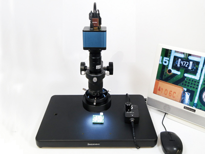
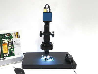
How to attach translucent light to microscope without light?

We will show you how to attach translucent light to the base without transillumination afterwards.

(There are some conditions based on base shape.)

透過照明

As mentioned above, there are the following methods as long as it is a microscope with an observation board (black and white plate).

Firstly, remove the observation board.

透過照明

Place the surface emitting light under the base. Pull out the cable from the gap.

透過照明

Attach the glass plate instead of the observation board.

顕微鏡

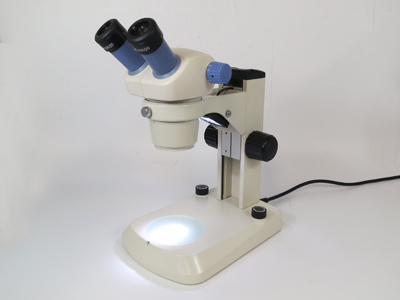
Adding a glass plate and surface emission lighting can be used as a simple transparent lighting stand.


透過照明   透過照明


<Transmitted lighting used>

透過照明   Light illumination for small camera stand RD-95T


RMS STANDARD

The RMS (Royal Microscopical Society) standard is the specification of the objective lens connection part (M20.32 mm P = 0.7.6 mm) of the microscope.


RMS → C mount

RMS → T mount


Conversion adapters such as are commercially available.

You can do various things by using this.

How to take a photograph by using microscope with a scale?

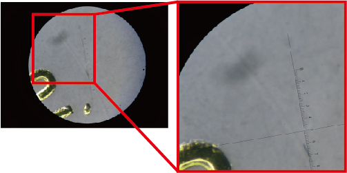
There are scales on the software.

The pitch of the scale can be changed

The above picture is adjusted to divide the 0.1 mm width into 25 equal parts. (You can not put numbers on the scale.)


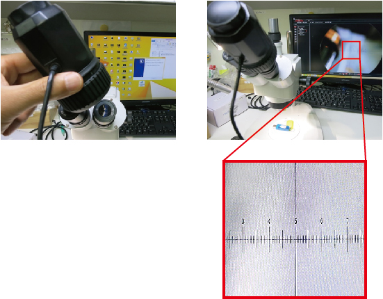
(1)Shooting through a reticle (glass scale) lens with a small digital camera


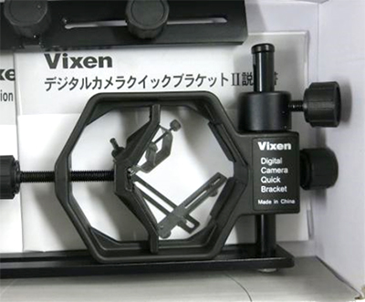
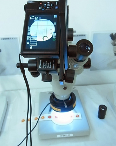
There is also a way to use ordinary digital camera and camera bracket.


Using this bracket, and attach a digital camera to the eyepiece of the microscope and shoot.


(2) Shooting with a microscope camera that covers the eyepiece


Place a eyepiece (glass scale) of the microscope.

Cover the eyepiece with a microscope camera that covers the eyepiece.


Then you can take pictures with a reticle (glass scale)

Tips for taking pictures with microscopes by single lens reflex camera

Focusing

Focusing during microscope shooting is not a microscope eyepiece but please go while watching the camera’s LCD screen. (It will be a viewfinder on cameras without live view function.)
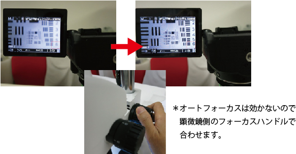
*auto focus function of camera doesn’t work at this time. so please adjust a focus with using focus angle of stereo microscope.


Even if you focus on an eyepiece, it usually does not match the focus of the camera.

(If there is diopter adjustment or back focus on the microscope, tonality may be possible.)

Vibration reduction

It will be like a picture blurred when you shake hands.

To prevent camera vibration, please use remote control or set the time settings.

For cameras with low vibration mode, set the low vibration mode to ON (about 1 second).

Exposure adjustment

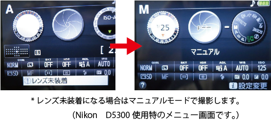
The exposure priority mode (dial A) is easy when shooting. If the monitor is too dark or too bright, please adjust the light / dark control (±).

Depending on the camera, “lens unmounted” may become occasionally.

In this case, shoot in manual mode.

(In this case, the brightness is adjusted by the shutter speed.)
White balance

Please manually adjust if the image becomes reddish or blue-colored due to the color of the light source of the microscope.

Make the light source of the microscope proper, remove the specimen, set it to nothing on the screen (glass part without specimen in the case of a biological microscope, white or gray board in case of a stereomicroscope), set the screen to white I will let you recognize.

※ Please see the operation manual of the camera for detailed operation.

ISO sensitivity

If the ISO sensitivity is too high, the image quality will degrade. If the screen is dark, you can take a beautiful image by using some method such as brightening the light of the microscope or slowing down the shutter speed, setting the sensitivity to a lower setting and shooting.

How to save a wide field of view with a camera of microscope

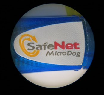
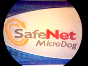
You can see the visual field of the naked eye observation of the microscope is the right picture   顕微鏡の裸眼観察の視野
     

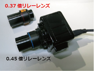
Normally, a 1/2 inch camera and a 0.45-megapixel lens will be included in the kit.

In this case, the field of view will be as shown on the right.

With a connector len of 0.45 times, vignetting (shadow) does not occur at the four corners of the image.

  0.45倍リレーレンズ
     

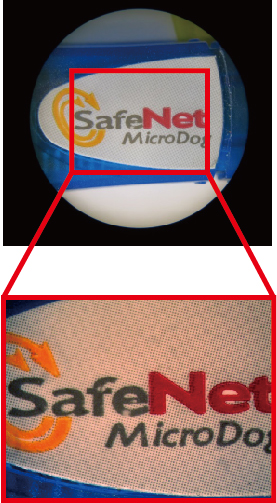
In case you really want to get a wide field of view, if you attach a wide 0.37x relay lens (wide angle) to the 1/2-inch camera, it will look like the right picture.

* Slightly, ambient distortion occurs.

  0.37倍リレーレンズ
     
    リレーレンズ

How to prevent reflection of lighting by binocular microscope

For monocular len microscope, there are indirect lighting devices such as dome lighting and arch lighting.

間接照明

However, for a binocular stereomicroscope, it is structurally two light path designs.

Therefore, the lighting device like the one above can not be used as it is.

間接照明

There are also a way to weaken the reflection by using the LED (flat type) with the LED pointing straight down as below.

間接照明

For simplicity, there is also a method to observe by attaching a diffusion tube to the LED ring light

間接照明

How to attach general-purpose LED ring light to stereo microscope?

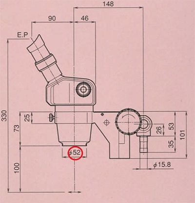
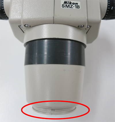
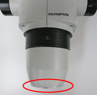
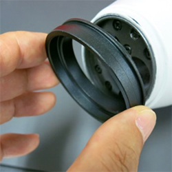
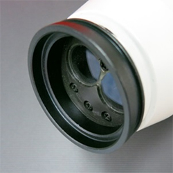
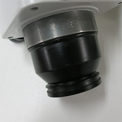
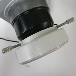
When attaching general-purpose LED ring light (not special LED ring light) to the tip of a stereo microscope, if the inner diameter of LED ring light is larger than the tip part of the microscope (the red circle part of the below picture), basically it can attach.


(Microscope with attachment part at the tip)


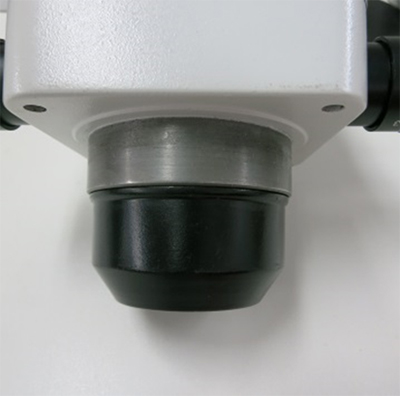
Based on the type ofmicroscope, there is a T-bar hanging at the head or the head is larger than the inner diameter of the LED ring light.


(Microscope without attachment part at tip)


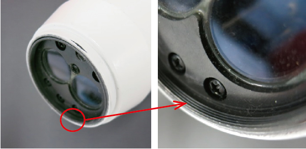
In such a case, you can easily attach the lighting by attaching the LED fixing ring using the inner screw of the microscope.

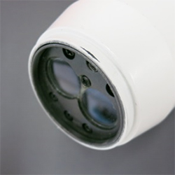
For microscope as described above, the inner screw for fixing the filter is cut on the back of the tip cover.

As it is for optical filters, pitch microscope is common regardless of microscope manufacturer. (P = 0.75 mm)

* By the way, it is a fine pitch than fine thread of he general ISO screw.


This can be used to attach the fixing ring.
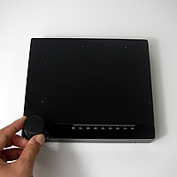
(We have a fixed ring of M49 and M48 available.)


It is easy to attach LED ring light.
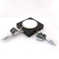


The inner thread diameter may be large with a very large microscope.

There is no problem in this case.

Step-down rings are on sale from filter makers and others.


The following example is a step down ring from M 58 to M 49 mm.


By combining an LED fixing ring (attach with step down ring M49), you can easily fix the LED ring light to a microscope with a large aperture.

WHAT’S XY TABLE?

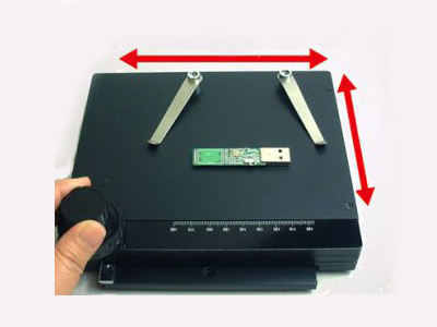
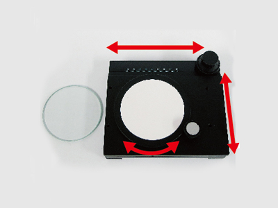
A support table (sample table) with a handle that can be moved in X and Y directions. It is advantageous when using the appropriate position adjustment becomes easier.

There are several methos to move the XY table.

1. Direct acting gear (that moves the XY table using the steering wheel)


簡易XYテーブル(落射照明タイプ)
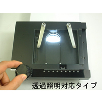
簡易XYテーブル(透過照明タイプ)

Simple XY table (Transparent light)

TK100-N


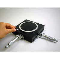
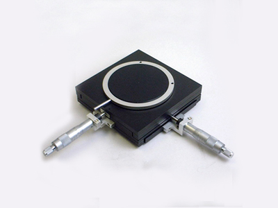
2. XY table using micrometers

You can move precisely. It is an advantage to be able to measure the amount of movement.

マイクロメータ付きXYテーブル
デジタルマイクロメータ付きXYテーブル

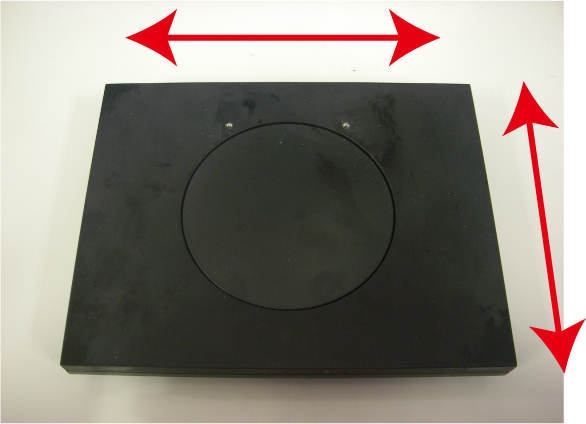
3.XY table without knob

Since the steering wheel does not protrude, it is possible to place big observed objects.

Sometimes it is called a slide table.

スライドテーブル(特注品) Sliding table
(Special order)


※Sliding table movement

LUNAR NEW YEAR HOLIDAY

Shodensha Vietnam is pleased to announce our schedule of Lunar New Year holiday as follow:
-Our office will be closed from Saturday,  10th February 2018 to Tuesday, 20rd Februry 2018
-Business operation will resume as normal on February 21th2018 (Wednesday).
 
We would like to take this opportunity to thank you for your continuous support.

HAPPY NEW YEAR HOLIDAY

Thank you for your trust, using our products and cooperate with us for a long time.
Shodensha Vietnam is pleased to announce our schedule of Happy New Year holiday as follow:
 
-Our office will be closed from Saturday,  30 th December 2017 to Wednesday, 3rd January 2018
-Business operation will resume as normal on Thursday 4th2018 (Tuesday).
 
Wishing you and your family a happy holiday.

DEFINITION OF MICROSCOPE MAGNIFICATION

The definition of microscope magnification is quite simple.
For eg: If the observed object (1mm) is displayed on the PC monitor or PC to 10mm, the magnification will be 10 times

Caculation:

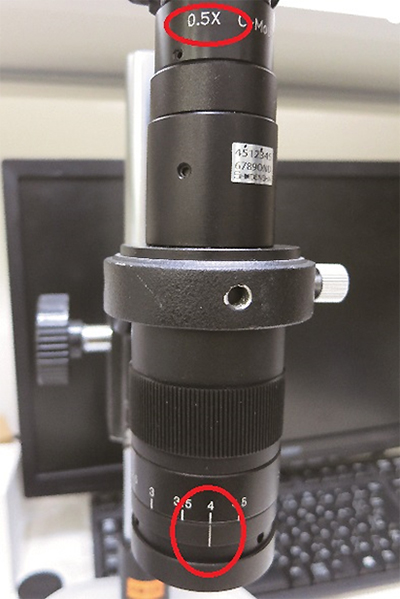
General magnification = monitor magnification x optical magnification (lens scale)

Monitor magnification = monitor size (inch) x 25.4mm/flange distance

Flange distance 6mm (1/3” camera), 7mm (1/2.5” camera), 8mm (1/2” camera) (Please refer to [Image size of camera] for more information)

The magnification of the lens was calculated value when it was attached to our 1/2 inch camera (variable magnification lens for coaxial lighting SDS-FZ)  and observed with a 17-inch monitor.

Monitor magnification = (17×25.4)/8 = 54

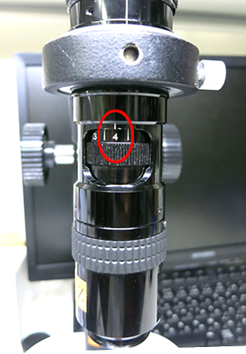
General magnification = 54×4 = 216 times

高解像度高倍率ズームレンズ SDS-FZR
   

By combining camera to auxiliary lens, ring lens, conveter lens, objective lens, …, the magnification will be increased.

The magnification of the lens was calculated value when it was attached to our 1/2 inch camera (Standard variable magnification lens SDS-M) and observed with a 17-inch monitor.

Monitor magnification = (17×25.4)/8 = 54

General magnification = 54x4x0.5 (lens magnification) = 108 times

ズームレンズ SDS-M


■ How to show filed of view of storaged images?

The magnification of the microscope has a lot of parameters, it becomes complicated when writing reports.

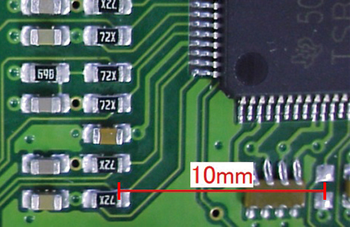
Therefore, please keep the calibration value instead of magnification that shown in the right picture.

Then you can determine the approximate field of view of the image.

計測ソフトMF Ship
   

Exhibition Announcement

Shodensha Vietnam is going to participate in an exhibition, Metalex Vietnam 2017,

held at Saigon Exhibition and Convention Center(SECC)on Oct. 12th – 14th.


We will hand over a 15% discount card※ and a small loupe in exchange for a business card.

※Except for some items. There is an expiration date.


◆◇◆INFO of Metalex Vietnam 2017◆◇◆

Name: Metalex Vietnam 2017 (Vietnam’s international exhibition on machining tools and metalworking solution for production upgrade)

Date: Oct. 12th – 14th 2017

Hours: 9AM – 5PM Daily

Place: Saigon Exhibition and Convention Center(SECC)

Booth: B 08 (Hall A)

NATIONAL HOLIDAY ANNOUNCEMENT

Thank you for your trust, using our products and cooperate with us for a long time.

Shodensha Vietnam is pleased to announce our schedule of Independence Day holiday 02nd September 2017 as follow:


-Our office will be closed from Saturday,  2ndSeptember 2017 to Monday, 4th September 2017

-Business operation will resume as normal on September 5th2017 (Tuesday).


Wishing you and your family a happy holiday.

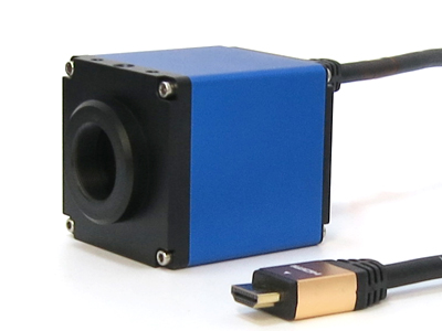
FULL HD Camera GR200HD2

A high defination camera with the resolution of 1920 x 1080.
It can be easily connected to a monitor with an HDMI cable.
※Please use a wide monitor of 16:9.

60fps smooth display and the high quality of the picture produced (5 times clear than video camera).

Images can be saved with Built in USB Flash slot(Image saved in BMP format)

* Please use a 32GB or below USB flash.

※Lens is optional.
HDMI cable comes as standard equipment.


※We recommend a PC monitor with an HDMI terminal, industrial monitor, etc.
(TV monitors are compatible but sometimes may not be shown.)
Demonstration products are available. For further information, please contact our technical support division.

●Relate to Monitor connect with HD Microscope→Check detail information here

Low price Stereo zoom Microscope NSZ-405

Low price zoom microscope! At this low price, it is highly efficient stereo microscope with full-scale specifications, having 7X – 30X zoom lens, epi-illumination, 2 pc of built-in LED light for transillumination.

  • Dust cover
  • LED light of epi-illumination and transillumination (with dimming)
  • Rubber eye-cups
  • Stage

Auto Focus Microscope HD TG200AHD1

A high definition Auto Focus Microscope with resolution of 1920 x 1080.

Available to observe it with a high-resolution image of the full high-definition.
Easily connect to monitor with HDMI cable.
※Please use a widescreen 16:9 monitor


  • Total magnification of 25 to 150 times enabled by an optical 6x zoom lens.( W.D. 90mm)
  • A zoom lens with latch function to lock each memory. (It is suitable to reduce error of measurement by the operator.)
  • A 0.5x auxiliary lens is offered (optional).
  • Resolution:1920×1080 (FULL HD)
  • No display delay(60fps)
  • Images can be saved with Built-in Micro SD card (Still image: JPEG format/ Movie: H.264 format)
  • Easily connect to monitor by HDMI Cable comes as standard equipment.

*Please use a 32GB or below USB memory

*We recommend a PC monitor with an HDMI terminal, industrial monitor, etc. 
(TV monitors are compatible but sometimes may not be shown.)
Demonstration products are available. For further information, please contact our technical support division.

Relate to Monitor connect with HD Microscope→Check detail information here

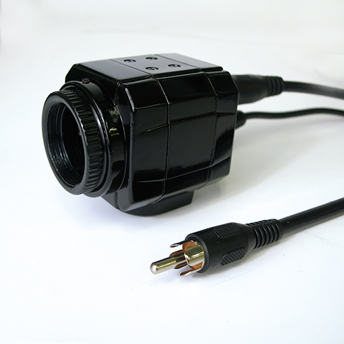
Video Camera GR-i700

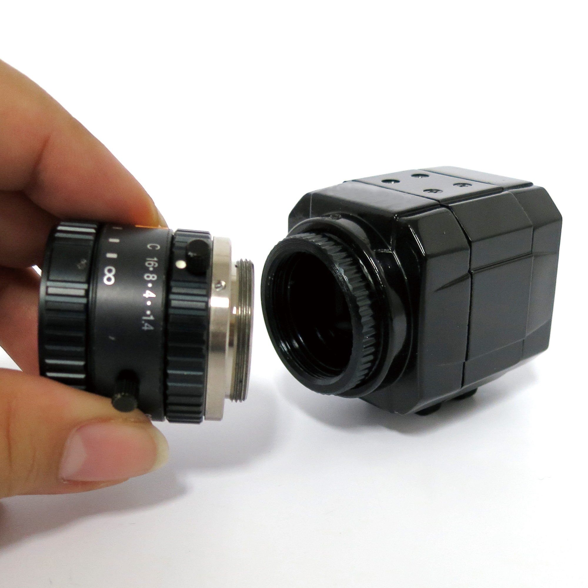
  • C Mount Camera
  • Achieving a smooth movie with clear image
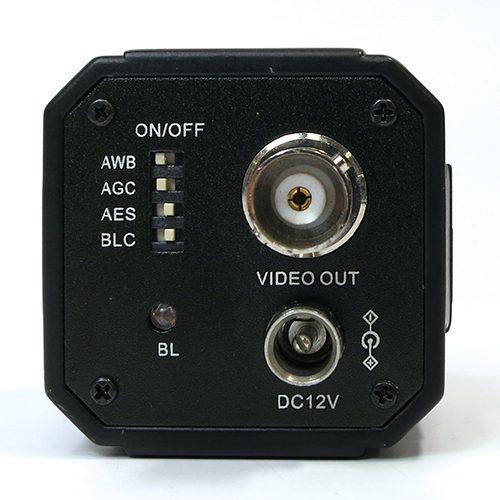
  • Available to adjust Auto White Balance and lock the setting
  • Available to select Auto exposure or manual exposure
  • NTSC Video Signal
  • BNC-RCA converter cable comes as standard equipment

GR-i700_cmount

<C Mount camera>


GR-i700_back2

Switchable ON/OFF of each function on the back of camera


High Magnification Zoom Trinocular Stereo microscope GR1040-65S3

Zoom Stereo Microscope with dimmable 56 LED Ring Light.
High magnification lens for wider observation field is adopted.
Available to observe from low magnification 10x to high magnification 65x with wide range.
Low price is achieved.
This trinocular stereo microscope has back focus function on the camera port.


Optional Auxiliary Lens

Tet Holiday Information

From Thu. 26 January 2016 to Wed. 1 February 2017,

we will be closed for Tet holiday (New Year Holiday).

We will be able to reply inquiries from Tue. 2nd of February.

Thank you and Happy Lunar New Year!

FULL HD Microscope TG200HD2

A high definition microscope with resolution of 1920 x 1080. (Approximately 5 times higher than the resolution of conventional video microscope.)
Available to observe it with a high-resolution image of the full high-definition.
Easily connect to monitor with HDMI cable.

※Please use a widescreen 16:9 monitor

・Total magnification of 25 to 150 times enabled by an optical 6x zoom lens.
・A zoom lens with latch function to lock each memory. (It is suitable to reduce error of measurement by the operator.)
・A 0.5x auxiliary lens is offered (optional).
・Working Distance : 90mm
・Resolution:1920×1028 FULL HD (1080p)
・No display delay(60fps)
・Images can be saved with Built-in USB memory slot (Image saved in BMP format)
・HDR (High Dynamic Range)function
・Display / Hide crossline(Black/White line)
・Connect to HDMI / Wide monitor with HDMI cable
・Built in external trigger terminal


※For HDMI, PC monitor or industrial monitor are preferred.
(TV monitors are compatible but sometimes may not be shown.)
Demonstration products are available. For further information, please contact our technical support division.

Relate to Monitor connect with HD Microscope→Check detail information here

Full HD Microscope With Built In Measurement Function TG200HD2-Me

An excellent performance of the 1080p microscope camera come with built-in measurement function.

・The microscope with built-in measurement function
・HDR (high dynamic range) function
・6x optical zoom lens with total zoom of 30x~180px
・Support 0.5x auxiliary lens (optional)
・W.D. : 90mm
・1920×1028 resolution (Full HD)
・High frame per second (60fps)
・Easy to save image (BMP) with it built in USB memory slot.
・Comes with an HDMI cable.


※For HDMI, PC monitor or industrial monitor are preferred.
(TV monitors are compatible but sometimes may not be shown.)

Demonstration products are available. For further information, please contact our technical support division. ●Relate to Monitor connect with HD Microscope→Check detail information here

Rich Function Full HD Microscope TG200HD2-MePRO

Measurement function, arbitrary cross line generation function, concentric circle generation function, angle line generation function have been added to the camera of Full HD microscope which can be observed with high quality images of full high vision (Full HD).

No PC Required! Available to Measure on the monitor. The Screen of measurement result can be saved in USB flash.
  • Measurement Function
  • HDR(High Dynamic Range)Function
  • Thumbnail display function
  • Total magnification of 30 to 180 times enabled by an optical 6x zoom lens.
  • A zoom lens with latch function to lock each memory. (It is suitable to reduce error of measurement by the operator.)
  • A 0.5x auxiliary lens is offered (optional).
  • Working Distance : 90mm
  • Resolution:1920×1028 FULL HD (1080p)
  • No display delay(60fps)
  • Images can be saved with Built-in USB Flash slot(Image saved in BMP format)
  • HDMI cable comes as standard equipment


1.3 Megapixel USB 3.0 Camera DN3RG-130

  • Machine vision camera using USB3.0.
  • Camera mount C(available for use as CS mounting by taking off a 5mm close-up photography ring)
  • Include outside trigger input terminal.
  • Strobe  output terminal is attached.
  • Global shutter model makes a lineup while being a CMOS sensor.
  • Prevent frame dropping function is attached so that frame rate will be prevented from becoming late by the affinity with the PC
  • The double setup of the same model is possible.
  • Saving image, video by Viewer software.
  • There are four holes of the M3 in the camera base so that can fix it well.
  • The tripod adapter is optional.
  • The USB3.0 cable (2m) with screw is optional

usb30_back 

<Back of camera>

Print

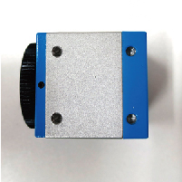
<Bottom of Camera>

2.0 Megapixel USB 3.0 Camera DN3RG-200

  • Machine vision camera using USB3.0.
  • Camera mount C(available for use as CS mounting by taking off a 5mm close-up photography ring)
  • Include outside trigger input terminal.
  • Strobe output terminal is attached.
  • Global shutter model makes a lineup while being a CMOS sensor.
  • Prevent frame dropping function is attached so that frame rate will be prevented from becoming late by the affinity with the PC
  • The double setup of the same model is possible.
  • Saving image, video by Viewer software.
  • There are four holes of the M3 in the camera base so that can fix it well.
  • The tripod adapter is optional.
  • The USB3.0 cable (2m) with screw is optional

usb30_back

<Back of camera>

Print

<Bottom of Camera>

5.0 Megapixel USB 3.0 Camera DN3R-500

  • Machine vision camera using USB3.0.
  • Camera mount C(available for use as CS mounting by taking off a 5mm close-up photography ring)
  • Include outside trigger input terminal.
  • Strobe  output terminal is attached.
  • Global shutter model makes a lineup while being a CMOS sensor.
  • Prevent frame dropping function is attached so that frame rate will be prevented from becoming late by the affinity with the PC
  • The double setup of the same model is possible.
  • Saving image, video by Viewer software.
  • There are 4 holes of the M3 in the camera base that can be used to fix camera well.
  • The tripod adapter is sold separately.
  • USB3.0 cable with screw is sold separately.


usb30_back 

<Backside of camera>


Print


<Camera base>

Holiday Information

From Sat. 31 December 2016 to Mon. 2 January 2017,

we will be closed for the New Year’s holiday.


We will be able to reply inquiries from Tue. 3th of January.


We apologize for the inconvenience and wish you a wonderful year.

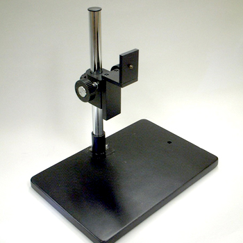
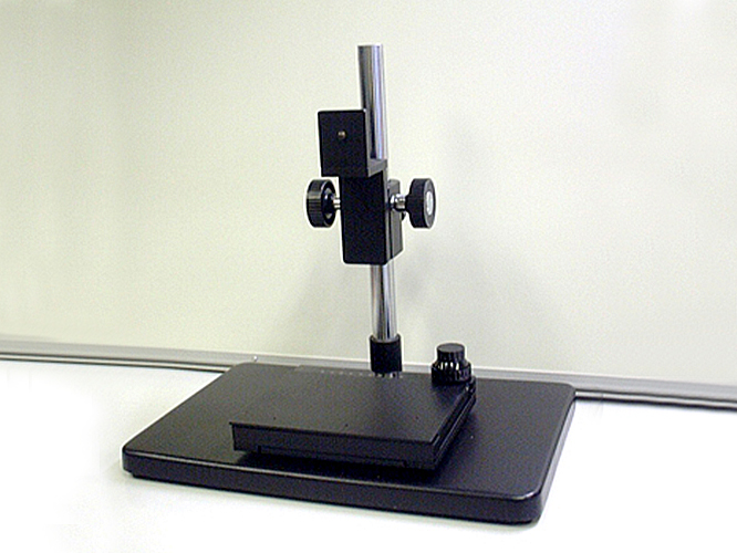
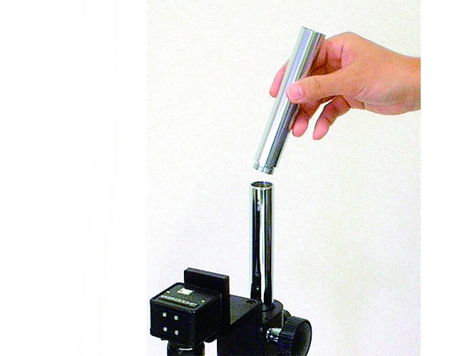
Extension Kit EK-135

You can extend the length of mounting post using it to fit to the object.
(It is usable for all of GR-STD1,GR-STD2,GR-STD11)

Extension length: 135mm per each, it is able to use maximum 3 pieces.

Shodensha Vietnam Co., Ltd. is established

Shodensha, Inc. established Our Vietnam Office in Ho chi minh, Vietnam.

Shodensha’s Optical products such as Digital Microscope and Stereo Microscope can be purchased in Vietnam.
Please do not hesitate to contact us.

Company Information
Shodensha Vietnam Co., Ltd.
(Công ty TNHH Shodensha Việt Nam)

Address
#1623, CJ Building, 2bis-4-6 Le Thanh Ton Street, Ben Nghe Ward,
District 1, Ho Chi Minh City, Vietnam

Tel Number 08-6255-6883
Fax Number 08-6255-6884
E-mail info@shodensha.com.vn (Japanese・Vietnamese・English Available)

10.0 Megapixel USB 3.0 Camera DN3R-1000

  • Machine vision camera using USB3.0.
  • Camera mount C(available for use as CS mounting by taking off a 5mm close-up photography ring)
  • Include outside trigger input terminal.
  • Strobe output terminal is attached.
  • Global shutter model makes a lineup while being a CMOS sensor.
  • Prevent frame dropping function is attached so that frame rate will be prevented from becoming late by the affinity with the PC
  • The double setup of the same model is possible.
  • Saving image, video by Viewer software.
  • There are 4 holes of the M3 in the camera base that can be used to fix camera well.
  • The tripod adapter is sold separately.
  • USB3.0 cable with screw is sold separately.


usb30_back

<Backside of camera>


Print


<Camera base>